‘Y Chromosome’- Adam & ‘Mitochondrial’- Eve
By Dr Zafar M. Iqbal
TCCI, Chicago, IL
When we think about human ancestry, the term scientists commonly use is ‘Most Recent Common Ancestors (MRCA). Describing them as ‘Y Chromosome’-Adam and ‘Mitochondrial’-Eve, as some have done, is not quite acceptable to many scientists, first, because it is misleading and, then, they’d rather not mix science with such faith-based references. May not be cuddly or scientific enough for them, but it does have the genetic flavor if you ignore the misleading Genesis connotations!
Early phylogenetic evidence first suggested that our ‘ancestor’ was probably a woman who lived in Africa 200,000 years ago (Cann, Stoneking & Wilson, Nature 325: 31-36, Jan 1, 1987; doi:10.1038/325031a0 ). In fact, that ancestor was more accurately the MRCA through matrilineal descent of all humans alive today.Some called her ‘Eve’ who, based on some other data, could have lived at least tens of thousands of years before Adam.
Who came first is not just another faith-based questions, it also continues to bother the anthropologists, palaeontologists and geneticists alike. A lot of information exists on how the modern man, Homo sapiens, evolved and lived first in Africa 200,000 years ago, and then spread to other continents over the centuries (the ‘out-of-Africa’ scenario).
Approaches to pursuing genetic genealogy are gender-based. Humans have 46 chromosomes (23 pairs) in each cell, including 2 sex-determining chromosomes, X and Y (females with XX and males XY). Y chromosome, with nearly 60 million DNA base pairs, is of particular interest because its DNA is passed on almost unchanged, from father to son. Testing Y-STR (Short Tandem Repeats) in Y-DNA is commonly used for determining patrilineal (or paternal) ancestry.
The other genetic genealogy tool uses mitochondrial (mt) DNA. While most (98%) of cell DNA is packaged and housed in the nucleus, a tiny amount (~2%) is found in the mitochondria. Each cell contains in its cytoplasm hundreds of thousands of mitochondria, cellular organelles which are involved mainly in converting food energy by oxidative phosphorylation, releasing ATP, the main source of energy for cells. Mitochondria also have a circular and double-stranded DNA, composed of 37 almost contiguous genes, separated by one or two bases, with nearly 17,000 nucleotide base pairs in each of 2-10 copies of the genetic material in each mt. Besides being tiny in size compared to its counterpart in the nucleus, mt DNA also has much higher mutation rate (10 -100 times) than nuclear DNA, lacks adequate DNA repair mechanism and, most important in genetic genealogy, mtDNA is inherited from mothers to her children, both male and female, which then is passed on to the next generation only by the female. During fertilization, both sperm and egg cells have mt DNA, but when sperm enters the egg, mtDNA of sperm is left outside the egg.
Both these genetic tools were used in two independent studies on MRCA, published in the prestigious American weekly ‘Science’ (August 2).
In one paper (Poznik et al: Science 341: 562-565; http://www.sciencemag.org/content/341/6145/565.abstract ), the Stanford researchers looked at complete Y-chromosome sequences among 69 men from nine global regions, including Pakistan. Other regions were Namibia, the Democratic Republic of Congo, Gabon, Algeria, Cambodia, Siberia and Mexico.They uncovered about 9,000 previously unknown DNA sequence variations, based on which they created a more reliable molecular clock. They obtained reliably accurate sequencing over a length of about 10 megabases of Y chromosome DNA for each of the subjects in the study, and estimated the annual mutation rate on the Y chromosome (one mutation roughly every 125 years) by calibrating it with a known event (settlement of the Americas 15,000 years ago). They also worked with the mtDNA data from the same individuals and got a separate estimate on MRCA.
During these studies they were able to identify about 11,000 variants among the sequences which enabled them to determine, with much better accuracy, phylogenetic associations and timelines among the sequences, which helped them to better re-configure MRCA timelines. Molecular clock is used to determine not only when two closely related populations split but also how distant cousins are related.
As Poznik emphasized that they now have “a more complete structure [of the evolution tree], including meaningful branch lengths.” They estimated the time to MRCA (for Y chromosome) to be 120,000 to 156,000 years, and for mtDNA-based MRCA , this time ranges from 99,000 to 148,000 years. This suggests for the first time, contrary to previous evidence, some possible overlap between male and female lineages.
In the other independent study in the same issue of ‘Science’ 341: 565-569, 2013 ( http://www.sciencemag.org/content/341/6145/565.abstract ), Francalacci et al of University of Sassari in Italy, reported results of their study of genetic variations in samples taken from 1204 Sardinian men, which showed 11,763 male-specific single nucleotide polymorphisms (MSY-SNPs). More than half of these MSY-SNPs never seen before. Based on a phylogenetic tree they created out of their data, their MRCA estimates ranged from 180,000 to 200,000 years.
Using more precise techniques and a larger set of data, t hese studies showed the narrowing of age difference in MRCAs of Y-Chromosome Adam and Mitochondrial Eve. Now, it seems possible that both “could have produced individuals who found each other in the same space and time,” showing “for the first time that they overlap,” as someone familiar with the studies commented.
This doesn’t mean that all the scientific controversies have been settled with regard to human ancestry or the critics silenced. But by narrowing the age difference in male and female MRCAs and suggesting a possible overlap between them, these studies provide a less-confusing picture of our genetic genealogy.
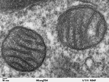
Fig 1: Two mitochondria from mammalian lung tissue displaying their matrix and membranes as shown by electron microscopy
http://upload.wikimedia.org/wikipedia/commons/thumb/0/0c/Mitochondria%2C_mammalian_lung_-_TEM.jpg/350px-Mitochondria%2C_mammalian_lung_-_TEM.jpg
-----------------------------------------------------------------------------

